Human Nerve Cell Labeled Diagram From the Ground
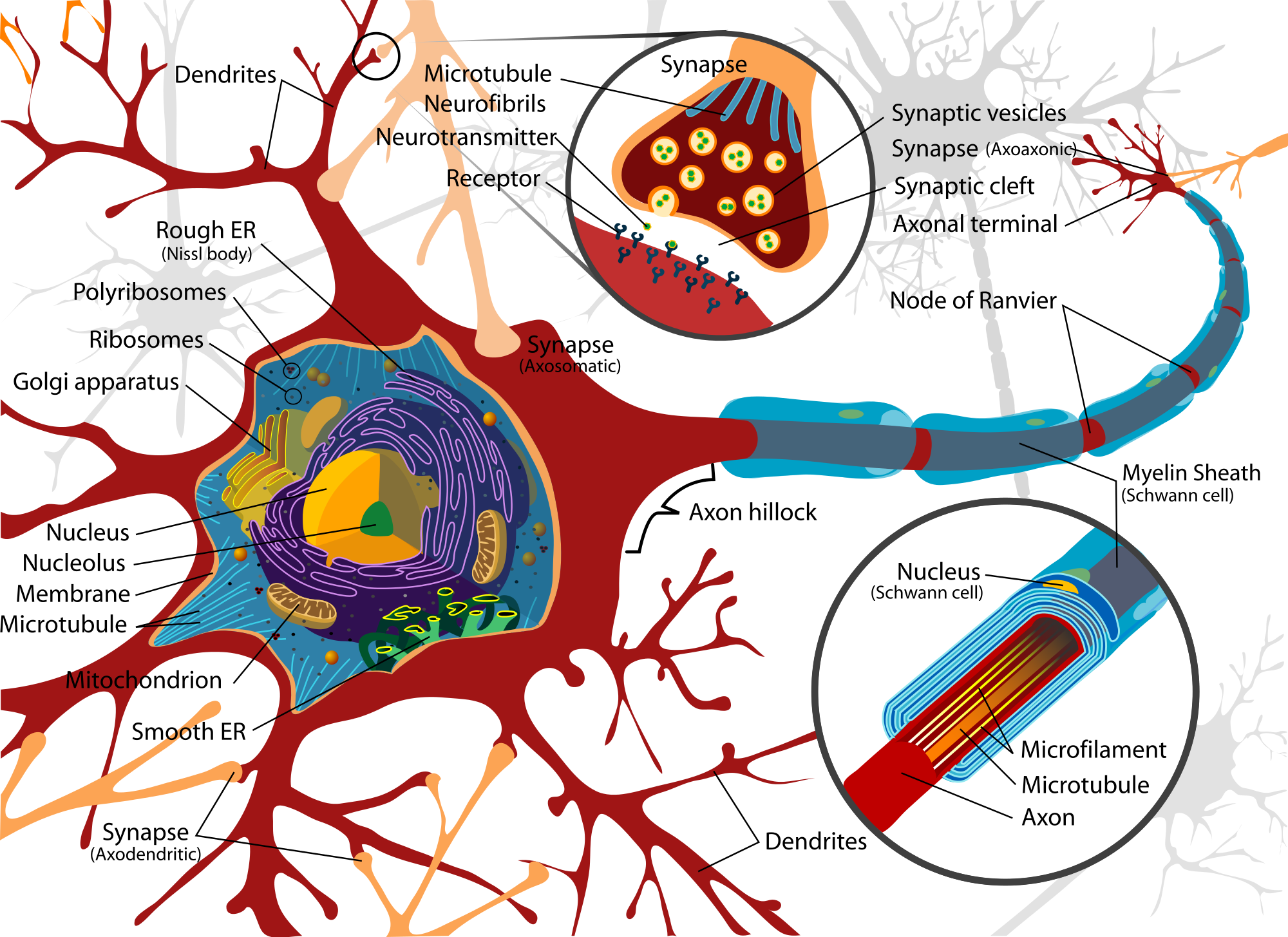
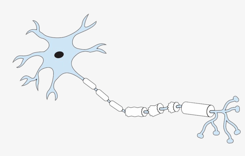
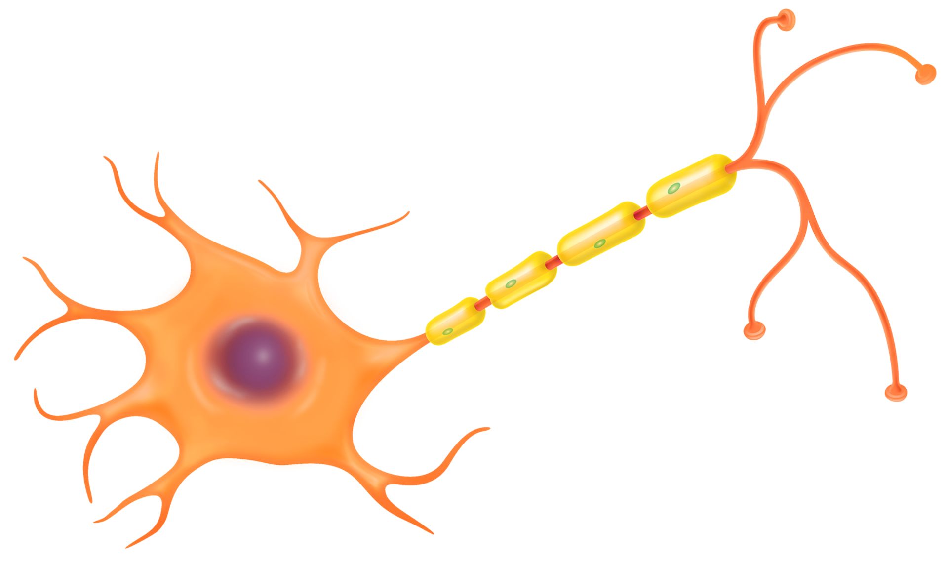
A neuron is a nerve cell that processes and transmits information through electrical and chemical signals in the nervous system. Neurons consist of a cell body, dendrites (which receive signals), and an axon (which sends signals). Synaptic connections allow communication between neurons, facilitating the relay of information throughout the body.

Neuron diagram, Neurons, Nerve cell
Diagram drawing and labelling of motor neuron for G.science and biology students

Image result for brain cell tattoo Tattoo Neurons, Brain, Neuroscience
Neurons (nerve cells) are the functional units of the nervous system. Even though they vary in size and shape, most have structural characteristics similar to the spinal cord neuron shown to left. Neurons have at their core an expanded area of cytoplasm called the cell body (soma or perikaryon). 1. 2.
Nerve Cell Diagram Labeled ClipArt Best
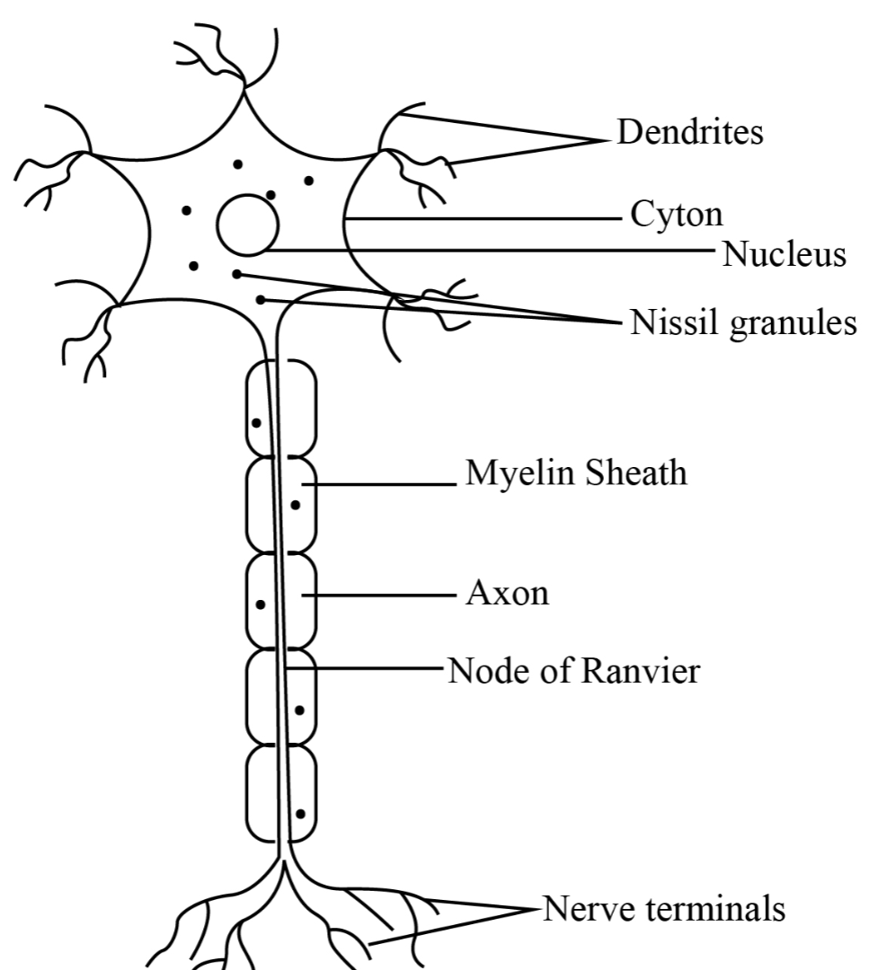
Parts of the Nerve Cell and Their Functions Silvia Helena Cardoso, PhD [1. Cell body] [2.Neuronal membrane][3.Dendrites] [4. Axon][5. Nerve ending] 1. Cell body The (soma) is the factory of the neuron. It produces all the proteins for the dendrites,axons and synaptic terminals and contains specialized organelles such asthe mitochondria, Golgi.
What Is A Nerve? Structure, Function, Types of Nerves, Nerve Disorders
Nerve cells are also called neurons . They are adapted to carry electrical impulses from one place to another. They feature: an axon - a single nerve fibre that carries nerve impulses away.

Draw neat and labelled diagram of nerve cell. Brainly.in
Draw a neuron and label its key histological and structural features; Explain the microscopic structure of a nerve fiber, including the structure of the myelin sheath and connective tissue layers; Identify the four types of glial cells, their structures, and their functions; Explain the general layout of the spinal cord, cerebrum, and.

Parts of a Nerve Cell Nerve cell, Nervous system, Nervous
Shannan Muskopf activity, anatomy, labeling, learning, myelin, nervous, neuroglia, neuron, practice, slides Drag and drop activity where students label an neuron, neuroglial cells, and the connection between two interacting nerves; presented on Google Slides.
Nerve Tissue SEER Training
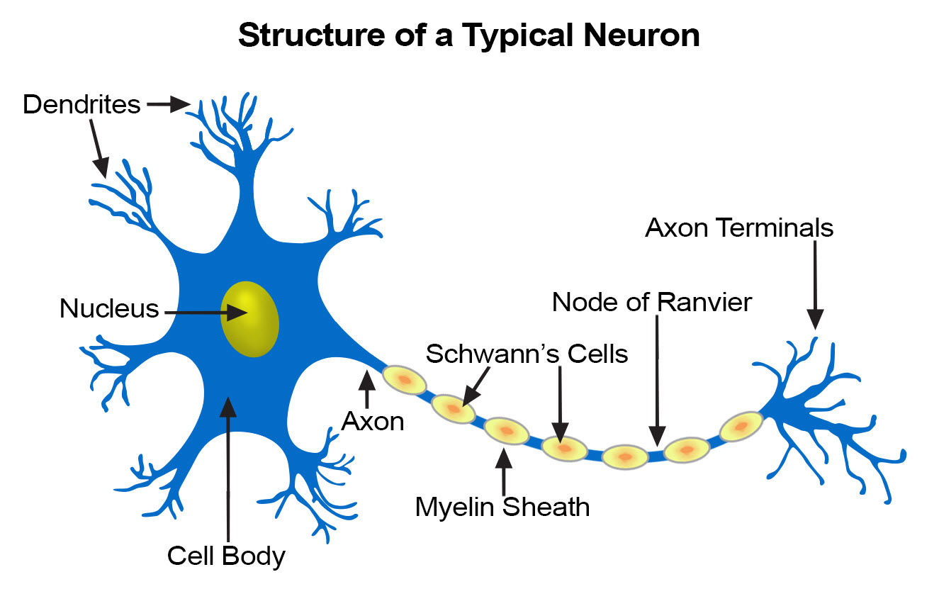
Most cells are 20 micrometers in diameter, which is just a fraction of the width of a hair. Neuron Anatomy Nerve Cell: Dendrites receive messages from other neurons. The message then moves through the axon to the other end of the neuron, then to the tips of the axon and then into the space between neurons.

Label The Neuron Answers
Nervous System - Neuron: Nerve Cell Name: Choose the correct names for the parts of the neuron. (1) (2) (3) (4) (5) (6) This neuron part receives messages from other neurons. (7) This neuron part sends on messages to other neurons. (8) This neuron part gives messages to muscle tissue. (9) This neuron part processes incoming messages.

Labeled Diagram Of The Neuron Stock Vector Illustration of image
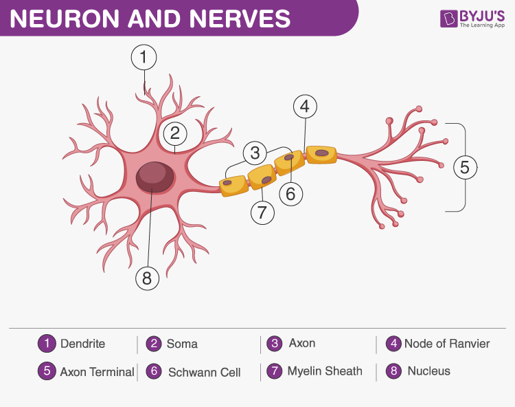
A nerve cell (neuron) consists of a large cell body and nerve fibers—one elongated extension (axon) for sending impulses and usually many branches (dendrites) for receiving impulses. The impulses from the axon cross a synapse (the junction between two nerve cells) to the dendrite of another cell.

Nerve Cell Diagram
The nervous system is a network of neurons whose main feature is to generate, modulate and transmit information between all the different parts of the human body. This property enables many important functions of the nervous system, such as regulation of vital body functions ( heartbeat, breathing, digestion), sensation and body movements.

Nervous System Anatomy and Physiology Nurseslabs Nervous system
Diagram Of Neuron with Labels Here is the description of human neuron along with the diagram of the neuron and their parts. The neuron is a specialized and individual cell, which is also known as the nerve cell. A group of neurons forms a nerve.

What is a nervous tissue? Give its functions. Explain the structure of
3,393 nerve cell diagram stock photos, 3D objects, vectors, and illustrations are available royalty-free. See nerve cell diagram stock video clips Filters All images Photos Vectors Illustrations 3D Objects Sort by Popular Education Chart of Biology for Nerve Cell Diagram. Vector illustration Basic Neuron Types Brain neuron symbol.

Brain Labeling Worksheet Biology worksheet, Cell diagram, Nerve cell
The central nervous system ( CNS) consists of the brain and the spinal cord. It is in the CNS that all of the analysis of information takes place. The peripheral nervous system ( PNS ), which consists of the neurons and parts of neurons found outside of the CNS, includes sensory neurons and motor neurons.
Human Nerve Cell Labeled Diagram From the Ground
An Easy Guide to Neuron Anatomy with Diagrams. Neurons, also known as nerve cells, send and receive signals from your brain. While neurons have a lot in common with other types of cells, they're.
Nerve Cell Function Nerve Cell Diagram DK Find Out
Click To View Large Image. The nervous system consists of the brain, spinal cord, sensory organs, and all of the nerves that connect these organs with the rest of the body. Together, these organs are responsible for the control of the body and communication among its parts. The brain and spinal cord form the control center known as the central.
